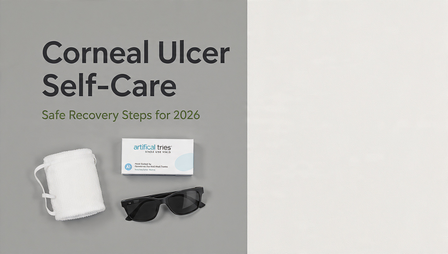
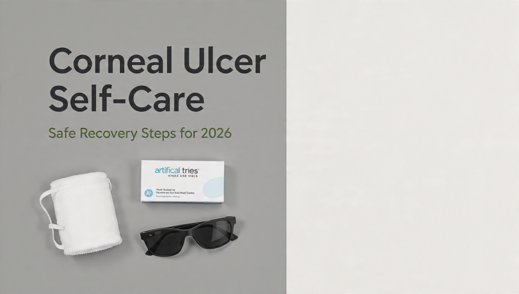
Corneal Ulcer Self-Care: Safe Recovery Steps for 2026
A corneal ulcer self-care routine is a doctor-prescribed, strictly supportive set of practices you follow at home while under active treatment from an ophthalmologist. It never replaces prescription antibiotic, antifungal, or antiviral eye drops. These self-care steps manage pain, protect the eye from further damage, and create an environment where the prescribed medication can work effectively.
The stakes are uniquely high with corneal infections. According to the Centers for Disease Control and Prevention (CDC), microbial keratitis, the clinical term for a corneal ulcer caused by infection, results in approximately 1 million outpatient and emergency visits annually in the United States. A corneal ulcer is an open sore on the clear dome covering the front of your eye. Damage here can lead to scarring and permanent vision loss if managed incorrectly. Your eye care specialist has given you a precise medical protocol. What you do between doses of that medication, in your daily habits, can either accelerate healing or create setbacks.
You will learn exactly how to apply a cool compress without pressure, how to wash your face without contaminating the eye, how to sleep in a way that protects the cornea, and how to recognize the early signs that your condition is worsening and requires immediate medical attention. You will learn the strict lines of what to avoid. And you will learn a safe timeline for reintroducing contact lenses, eye makeup, and facial skincare products after the ulcer has fully healed.
corneal ulcer self-care at home
Corneal ulcer self-care at home is a physician-directed set of environmental, hygiene, and comfort practices that support the healing of an open corneal wound. It is not a treatment for the infection itself. The American Academy of Ophthalmology (AAO) is explicit: antimicrobial therapy prescribed by an ophthalmologist is the only first-line treatment for infectious keratitis. Self-care is the safety scaffolding around that treatment.
Your primary goal at home is to protect the epithelial defect, the hole in the outermost layer of the cornea. Every blink, every drop of a non-sterile liquid, every unintentional rub of the eye mechanically disrupts migrating epithelial cells that are trying to close the wound. Your self-care practice creates a stable environment for those cells to bridge the gap. The second goal is to manage inflammation and pain without interfering with the therapeutic agents your doctor has prescribed.
Effective self-care breaks down into three immediate zones. First, physical protection of the globe to prevent further tissue loss. Second, strict avoidance of anything that introduces pathogens or toxic chemicals to the compromised corneal surface. Third, sensory management to control the intense photophobia (light sensitivity) and epiphora (reflex tearing) that accompany most corneal ulcers. Each of these zones requires precise, informed action, detailed in the sections ahead.
corneal ulcer immediate first steps
The immediate first steps after a corneal ulcer diagnosis are to stop wearing contact lenses, do not touch or rub the eye, begin the prescribed antibiotic or antifungal drop schedule immediately, and place any protective eye shield provided by your doctor over the eye. These steps are time-sensitive. Delaying the first dose of a prescribed fourth-generation fluoroquinolone like moxifloxacin 0.5% or a fortified antibiotic by even a few hours allows bacterial replication to continue at the epithelial wound margin.
Remove your contact lenses now. If you were wearing them when diagnosed, you have already been instructed to remove them. Do not store them in a case to reuse. They must be discarded along with the case they were stored in. The CDC contact lens health guidance states that microbial biofilms on lens cases are a primary vector for reinfection. Place the prescribed drops in your eye exactly as instructed. If you have both an antibiotic drop and a cycloplegic drop, wait at least five minutes between them to prevent the second drop from washing the first away.
If you were not given a rigid eye shield, an improvised one is better than none. The immediate priority is preventing any pressure on the globe during sleep or an inadvertent moment of rubbing. Tape a small, clean paper cup with the bottom cut out over the eye if a proper shield is unavailable. Secure it with medical tape to the bony orbital rim, not the soft eyelid. This prevents the shield from pressing into the eye. Call your ophthalmologist’s office and confirm you have a follow-up slit-lamp examination scheduled within 24 to 48 hours. Corneal ulcers can worsen within hours, and the initial culture results may demand a change in therapy.
cool compress for corneal ulcer pain
A cool compress for corneal ulcer pain is a damp, clean cloth applied lightly over the closed eyelid to provide temporary relief through vasoconstriction and reduced nerve conduction velocity. The mechanism is localized physical cooling. A 2022 review on corneal pain management published in the journal Cornea notes that cooling the periorbital skin reduces nociceptive firing in the trigeminal nerve branches that innervate the cornea. The pain signal is literally slowed down.
To apply a cool compress safely, wash your hands with antimicrobial soap for a full 20 seconds. Use a clean washcloth, not a reused hand towel. Wet it with cool tap water or chilled distilled water. The temperature should be cool, not ice-cold. Extreme cold can cause a rebound vascular dilation and damage the already fragile periocular skin. Wring it out fully. A dripping cloth introduces unsterile water that can migrate into the eye. Lay the damp cloth lightly over the closed eyelid. Do not apply any weight. The cloth should rest without pressing the eyelid into the cornea. Hold it there for 10 minutes. You can repeat this every two to four hours during the first 48 hours of acute pain.
Do not use gel eye masks, even if they are kept in the refrigerator. Their weight and the potential for a seal around the eye socket create pressure. Do not use ice packs. The periocular skin is thin and lacks the subcutaneous fat insulation of other facial areas, making it vulnerable to cold injury. Do not use frozen vegetables. They are not sterile and are a contamination risk. The cooled cloth is the only compress method with an acceptable safety profile for an open epithelial wound.
eye shield protection for corneal ulcer
An eye shield for a corneal ulcer is a rigid, ventilated plastic or metal dome placed over the eye to prevent mechanical pressure on the compromised cornea during sleep or rest. The eye shield is not an eye patch. The distinction is critical. A soft fabric patch can abrade the cornea, trap moisture, and create a dark, warm environment that may promote fungal growth if the ulcer is fungal in origin.
The eye shield must rest on the bony orbital rim, the hard bone of your brow and cheek, not on the eyelid. When taped correctly, the shield forms a protective arch over the eye. There is no contact between the shield and any part of the eyelid or cornea. This allows you to blink, apply drops through the holes, and allow tear film evaporation without obstruction. Secure it with three strips of medical-grade paper tape. Place one strip vertically across the center of the shield, and one on each side at a slight angle. Press the tape firmly onto the cheek, forehead, and the bridge of the nose. If you have a known allergy to adhesive tape, request a silicone-backed tape from your ophthalmologist.
Wear the shield every time you sleep, including daytime naps, until your ophthalmologist confirms the epithelial defect has fully closed under fluorescein staining. A closed eye during REM sleep can undergo rapid movements and Bell’s phenomenon does not always protect a centrally located ulcer. The shield is a mechanical insurance policy against a reflexive rub that erases days of epithelial healing in seconds.
how to manage light sensitivity from corneal ulcer
Light sensitivity from a corneal ulcer, clinically termed photophobia, is managed by dimming ambient light, wearing dark sunglasses indoors, and using the prescribed cycloplegic eye drop. Photophobia during a corneal ulcer is not a subjective preference for darkness. It is a neurobiological pain response triggered when light stimulates the intrinsically photosensitive retinal ganglion cells that converge on the trigeminal nerve pathway already sensitized by corneal inflammation. Light itself becomes a pain signal.
Your ophthalmologist will likely have prescribed a cycloplegic drop such as cyclopentolate 1% or atropine 1%. This medication temporarily paralyzes the ciliary muscle and dilates the pupil. The paralyzed ciliary muscle stops the painful spasm of accommodation that occurs when the eye tries to focus. By dilating the pupil, it also reduces the light entering the eye. Do not skip this drop. Even if the dilation makes near vision blurry, it is a critical component of pain control during the first 48 to 72 hours.
In your home, draw the curtains. Lower the brightness on every screen. Most phones and laptops have an accessibility setting to reduce white point, which is more effective than standard brightness reduction. Wear dark, wraparound sunglasses indoors. Sunglasses with complete peripheral coverage block ambient light from entering from the sides, which is especially triggering for photophobic pain. At night, use a dim nightlight in the bathroom rather than switching on the overhead light for middle-of-the-night drop applications. Sudden brightness triggers a disproportionate pain spike.
what to avoid doing with a corneal ulcer
What to avoid doing with a corneal ulcer includes wearing contact lenses, applying any unprescribed eye drop, rubbing the eye, swimming, showering without a shield, and using any facial product that can migrate into the tear film. The list of prohibitions is strict because the open wound on the cornea has zero barrier function against pathogens and chemicals.
Contact lenses are a wound contaminant. Do not reinsert them until your ophthalmologist has cleared you, which typically requires a fully closed epithelium and a period of stability. Applying a non-prescribed, over-the-counter redness relief drop that contains a vasoconstrictor like naphazoline or tetrahydrozoline can mask worsening inflammation and introduces the preservative benzalkonium chloride (BAK). BAK is a known corneal epithelial toxin. A 2020 study in the Journal of Ophthalmic Inflammation and Infection documented that BAK exposure to a compromised corneal epithelium increases stromal cell apoptosis. You cannot put anything into your eye that did not come from a prescription bottle.
Do not rub the eye. Tape the shield on before you sleep, tie a loose scarf around your hand, or wear a cotton glove on the hand of the affected side if you know you tend to rub your face in your sleep. Do not go swimming, sit in a hot tub, or submerge your head in a bath. Acanthamoeba, a free-living amoeba found in tap water, lakes, and hot tubs, causes a particularly devastating and treatment-resistant corneal ulcer. Do not apply makeup, eye cream, face oil, or sunscreen near the orbital bone. These substances migrate. Any product that can slide, melt, or drip into the tear film represents a foreign body risk and a microbial growth medium.
face washing safely with a corneal ulcer
Face washing safely with a corneal ulcer means cleaning the skin using a downward-only, no-splash technique that prevents any water, cleanser, or residue from entering the eye. You cannot splash water on your face. The corneal epithelium has a breach. Tap water is not sterile and can contain Acanthamoeba cysts, Pseudomonas, and other organisms that cause severe polymicrobial keratitis.
To wash your face, begin by removing any shirt that must be pulled over the head to avoid contact with the eye. Use a gentle, fragrance-free, non-foaming cleanser. Foaming cleansers contain surfactants like sodium lauryl sulfate (SLS) that can strip the skin barrier and, if they migrate into the tear film, cause significant stinging and epithelial irritation. A cream or lotion cleanser with ingredients like ceramide NP and glycerin is appropriate. Pump the cleanser onto a clean cotton round or your fingertips. Apply it to the non-affected side of the face, the forehead, the nose, and the chin, carefully avoiding the entire orbital area.
The jaw and neck: tilt your head downward and use a damp, clean washcloth to wipe the cleanser from the face in a downward motion only, from the forehead toward the chin. Never wipe upward. The upward motion would push product toward the eye. Rinse the cloth completely between each pass. For the skin under the affected eye, skip cleanser entirely if there is any epiphora, or continuous tearing. The constant overflow of reflex tears already cleanses this area; adding cleanser creates a slurry that can seep into the eye. Instead, use a clean tissue to gently blot the tearing from the outer canthus outward, never wiping toward the nose. Pat the face dry without rubbing.
sleeping safely with a corneal ulcer
Sleeping safely with a corneal ulcer requires you to sleep on your back or on the side opposite the affected eye, with the rigid eye shield securely taped in place. Your sleeping position directly affects intraocular pressure and the physical safety of the healing epithelium. Sleeping on the side of the affected eye presses the cornea against the pillow, a surface that is not sterile, and puts direct pressure on the globe. Sleeping face-down is unsafe.
Back sleeping is ideal. It keeps the eye facing upward, minimizes any pressure on the globe, and prevents the shield from being dislodged. If you are not a habitual back sleeper, construct a pillow barricade to prevent turning. Place a firm pillow on either side of your body and one under your knees. This configuration makes it physically difficult to roll onto your side during deep sleep. A travel pillow positioned around the neck can also discourage head turning.
The ocular surface heals during sustained closed-eye rest. The eyelid provides a protected, moist environment where epithelial cell migration and adhesion can occur without the shear force of blinking. The corneal epithelium regenerates fastest during uninterrupted sleep cycles. Prioritize sleep consistency. The eye shield tape must be checked before bed to ensure the edges have not lifted. A loose shield is a hazard. If the tape irritates your skin, apply a thin layer of a petrolatum-based ointment, like petroleum jelly, to the skin before taping, ensuring none migrates near the eye opening. The petrolatum acts as a protective interface between the adhesive and the stratum corneum.
Key Takeaway: Your sleep position and the rigid eye shield are your most important tools for preventing a mechanical setback. A single face-down or rubbing episode during REM sleep can undo days of epithelial healing.
corneal ulcer emergency signs and when to call a doctor
Corneal ulcer emergency signs include a sudden increase in pain, a visible white or yellow spot on the cornea that is growing, vision that has suddenly worsened, or the appearance of a fluid level of pus inside the eye known as a hypopyon. These are not signs to watch and wait with. These are signs to call your ophthalmologist’s emergency line immediately or go directly to an emergency department with ophthalmology coverage.
Increased pain after 48 hours of compliant antibiotic therapy is a red flag. Bacterial ulcers typically begin to feel less painful within one to two days of appropriate treatment. If the pain intensifies, the infection may be resistant to the prescribed antibiotic, or the ulcer may be extending deeper into the corneal stroma. Stromal melting, where collagen breaks down under the ulcer, is a vision-threatening progression. A visible increase in the size of the corneal infiltrate, the white spot you can see in the mirror, is objective evidence of progression.
A hypopyon, a yellowish-white layer of inflammatory cells settled at the bottom of the anterior chamber, looks like a tiny fluid level in the colored part of the eye. This finding means the inflammation has breached the cornea and entered the intraocular space. It requires immediate reevaluation and possibly a shift to fortified, compounded antibiotics or even a corneal culture with sensitivity testing. Sudden vision loss that is not explained by the blurred vision from your dilating drops is also an emergency. If the eye becomes suddenly softer or harder to the touch than the unaffected eye, this suggests a potential corneal perforation or acute glaucoma.
corneal ulcer healing timeline and recovery
The corneal ulcer healing timeline depends on the infectious organism, the size of the epithelial defect, and your underlying ocular surface health, but small, central, uncomplicated bacterial ulcers treated promptly can re-epithelialize within three to five days under aggressive antibiotic therapy. A corneal ulcer is considered healed when the epithelial defect is fully closed and no longer stains with fluorescein dye under slit-lamp examination by your ophthalmologist.
The re-epithelialization phase is visible to your doctor. Epithelial cells from the limbus, the border between the cornea and the white sclera, migrate centripetally toward the defect. They slide across the stroma like a sheet being pulled over a table. Over the first 48 to 72 hours, the defect size should measurably shrink. The pain of an epithelial defect, which is the sharp, sand-in-the-eye foreign body sensation, diminishes in direct proportion to wound closure. When the epithelium is sealed, the sharp pain resolves, though a dull ache may persist from residual stromal inflammation.
Stromal healing is a much longer, invisible process. Underneath the new epithelium, the corneal stroma remodels over weeks to months. The collagen fibers laid down during this phase are not always organized with the perfect parallel alignment of native cornea. This disorganized collagen can result in a corneal scar, a permanent opacity. Topical corticosteroids are sometimes prescribed after the epithelium has closed, specifically to modulate this scarring response, but only if the infection is confirmed to be bacterial or sterile and not herpes simplex virus or fungal. The final recovery endpoint, with stabilization of vision and scar density, can take six months or longer.
reintroducing contact lenses after a corneal ulcer
Reintroducing contact lenses after a corneal ulcer is a process that must be directed and timed by your ophthalmologist, not by your own comfort level, and typically requires a new lens prescription, a new case, and a strict switch to daily disposable lenses if you were using monthly or extended-wear lenses. The epithelial barrier and the nerve density of the cornea take far longer to normalize than the eye feels.
Your ophthalmologist will likely recommend waiting until all topical medications, including antibiotic drops and any post-closure corticosteroid drops, have been discontinued. The cornea must be completely quiet, with no residual punctate epithelial staining. A corneal topography or anterior segment optical coherence tomography (OCT) may be performed to ensure the stromal bed is of adequate thickness and shape to safely support a contact lens. The lens material matters. Silicone hydrogel daily disposable lenses are the standard recommendation post-infection because they eliminate the lens case, a known reservoir for biofilm-forming pathogens.
The lens fitting is a medical event, not a cosmetic refill. The curvature of the cornea can change subtly after an ulcer heals, especially if any stromal thinning occurred. Your old prescription will likely be incorrect and the base curve of your old lenses may no longer fit properly. A poorly fitting lens creates mechanical friction points on the newly healed epithelium. This is a setup for a recurrent erosion. Do not sleep in contact lenses after a corneal ulcer, ever. Even if a lens is FDA-approved for extended wear, the post-ulcer cornea carries a permanently elevated risk profile for infection.
eye makeup after corneal ulcer safety
Eye makeup after corneal ulcer safety protocols require you to discard all previously used eye makeup and applicators, wait until the ophthalmologist confirms the cornea is fully re-epithelialized and off all topical medication, and then reintroduce only new, preservative-free, ophthalmologist-approved products. The inside of a mascara tube, an eyeliner pencil, or an eyeshadow palette that was used before or during the infection is a contaminated item.
Bacteria, particularly Staphylococcus species, can survive in the waxes and oils of eye makeup. The FDA does not require cosmetics to be sterile, and the preservatives in cosmetics are designed for in-use contamination from a healthy microbiome, not to suppress a pathogenic load from an active infection. Discard every eye product you used in the two weeks before the ulcer diagnosis. This includes mascara, eyeliner, eyeshadow, eye primer, lash serum, and any brush or sponge that touched your eye area. Do not attempt to sanitize them. Sterilization of porous cosmetic products in a home setting is not possible.
Your ophthalmologist will likely recommend waiting at least one to two weeks after the epithelium has sealed and all medication has stopped. The waiting period is for the lipid layer of the tear film to stabilize and for the meibomian glands along the lid margin, which may have been inflamed, to return to normal function. When you are cleared, purchase new, fragrance-free, hypoallergenic eye makeup. Do not apply any product to the waterline. The waterline is the mucosal surface of the lid margin where the meibomian glands secrete oil. Blocking it with eyeliner introduces a particle load directly onto the tear film. A powder eyeshadow applied only to the mobile lid above the crease is the safest starting point.
periocular skincare to avoid with an eye infection
Periocular skincare products to avoid with an eye infection include any product containing chemical exfoliants, retinoids, essential oils, or physical exfoliating particles that could migrate into the tear film and cause chemical injury to the denuded corneal epithelium. The skin around the eye is thin and the distance from the orbital bone to the cornea is a matter of millimeters for a migrating cream.
Stop using any product containing alpha-hydroxy acids (AHAs) like glycolic acid or lactic acid, beta-hydroxy acids (BHAs) like salicylic acid, or retinoids including retinol, retinaldehyde, and prescription tretinoin. These compounds accelerate cellular turnover and thin the stratum corneum, which on healthy skin is desirable. Near an open corneal wound, contact with even a trace amount is a chemical insult. A 2019 review in the Journal of Investigative Dermatology confirms that retinoids increase corneal epithelial permeability in animal models. Do not apply these products anywhere on the face while the corneal epithelium is open.
Do not use any facial oil, cleansing balm, or heavy occlusive moisturizer near the eye. These products have a low melting point and will travel across the skin’s surface with body heat. Once in the tear film, oil creates an emulsion that blurs vision and, more importantly, can trap bacteria against the cornea. The only product that should be applied to the periocular skin during an active ulcer is a sterile, preservative-free ophthalmic ointment prescribed by your doctor. After the ulcer has healed, slowly reintroduce skincare products, starting with a simple ceramide moisturizer applied only to the cheekbone and temple, avoiding the orbital bone until you are fully cleared.
Key Takeaway: The cornea has no tolerance for cosmetic chemistry. During an active ulcer, the only product allowed on the skin adjacent to the eye is the ointment your ophthalmologist prescribes. All exfoliants, retinoids, and facial oils stop today.
Frequently Asked Questions About Corneal Ulcer Self-Care
Can I shower with a corneal ulcer?
You can shower with a corneal ulcer only if you wear a rigid eye shield securely taped to the orbital bone and keep your face completely out of the shower stream.
Tap water carries Acanthamoeba cysts and other organisms that can cause severe treatment-resistant corneal infections.
Tilt your head backward so water runs off the back of your head, never down your face.
How long does it take for a corneal ulcer to stop hurting?
The sharp, foreign-body sensation pain of an uncomplicated bacterial corneal ulcer typically decreases noticeably within 24 to 48 hours of starting effective antibiotic drops.
This pain relief correlates with the healing epithelium covering the exposed nerve endings.
A deep, aching pain may persist longer due to stromal inflammation and will resolve more slowly.
Can I watch TV or use my phone with a corneal ulcer?
You can watch TV or use a phone with a corneal ulcer, but you must reduce screen brightness to the lowest comfortable level and use a blue light filter or night mode.
Photophobia during a corneal ulcer is neurological pain triggered by light, not eye strain.
Wearing dark sunglasses while looking at screens and taking frequent blink breaks can make screen use tolerable.
What happens if water gets in my eye with a corneal ulcer?
If tap water gets into your eye with a corneal ulcer, immediately flush the eye with the sterile, preservative-free artificial tears you have been instructed to use and call your ophthalmologist’s office.
Tell them what happened and ask if you need an earlier re-evaluation.
Tap water is not sterile and introduces a risk of secondary polymicrobial or Acanthamoeba infection to the open wound.
Can I use over-the-counter eye drops for a corneal ulcer?
You cannot use over-the-counter eye drops for a corneal ulcer unless your ophthalmologist has specifically instructed you to use a particular brand of preservative-free artificial tear.
Redness-relieving drops contain vasoconstrictors and the corneal epithelial toxin benzalkonium chloride (BAK).
Do not place anything in the eye that did not come from your ophthalmologist’s prescription.
How do I know if my corneal ulcer is getting worse?
Your corneal ulcer is getting worse if the pain intensifies, the white spot you can see on the cornea grows larger, your vision drops suddenly, or you see a fluid level of pus in the lower part of your iris.
These are signs of infection progression or stromal melting.
Any one of these symptoms means you need to see your ophthalmologist or go to an emergency department immediately.
The line between a corneal ulcer that heals with a faint scar and one that results in permanent vision loss often comes down to the strictness of the safety practices you follow at home. Your ophthalmologist’s medication kills the pathogen. Your self-care prevents a second hit, a mechanical rub, a water-borne contaminant, or a cosmetic chemical from destroying the delicate cellular repair work that happens hour by hour. You are the guardian of the healing environment.
Tonight, tape on the eye shield. Check your side-sleeping barricade. Pull the curtains against the morning sun. And in the morning, wash your face with downward-only, careful hands. Every single one of these small, deliberate acts is a stitch in the wound you cannot see but that your ophthalmologist can measure in millimeters of closure.
This protocol of protection is temporary but absolute. The cornea has an extraordinary capacity to heal cleanly if it is given an uninterrupted chance. Give it that chance, one careful hour at a time.