Sleep Apnea Self-Care: What Actually Works in 2026
Sleep apnea self-care refers to the specific lifestyle and behavioral practices that reduce the frequency and severity of apnea events, improve sleep quality, and support any prescribed medical treatment a person is already receiving. These are not replacements for a CPAP machine or an oral appliance when those treatments are clinically indicated. They are the evidence-backed layer of daily habits that make a measurable difference in how well you breathe at night.
According to the NIH National Heart Lung and Blood Institute (NHLBI), obstructive sleep apnea affects an estimated 30 million adults in the United States, yet fewer than 20% of cases are currently diagnosed and treated. That gap matters because untreated OSA is independently associated with hypertension, cardiac arrhythmia, and type 2 diabetes. The self-care decisions you make every day directly affect how aggressively those risks compound.
This article covers the physiology of why OSA happens, which self-care interventions have randomized controlled trial support, what the CPAP-skin interface does to your skin and how to address it, and precisely when behavioral self-care stops being enough and professional escalation becomes necessary.
Sleep Apnea Self-Care: What It Can and Cannot Do
Sleep apnea self-care can reduce apnea frequency, lower the apnea-hypopnea index (AHI), and improve daytime symptoms, but its effectiveness depends entirely on OSA severity and anatomical factors that no lifestyle change can fully override.
Self-care is most impactful for people with mild OSA (AHI of 5 to 14 events per hour) or positional OSA (apneas that occur predominantly in the supine, or back-sleeping, position). In these groups, behavioral modifications can reduce AHI to below the diagnostic threshold in some individuals. For moderate OSA (AHI 15 to 29) and severe OSA (AHI 30 or above), self-care remains valuable but must accompany prescribed therapy, not substitute for it.
The American Academy of Sleep Medicine (AASM) classifies continuous positive airway pressure (CPAP) therapy as the first-line treatment for moderate and severe OSA. No lifestyle intervention has equivalent efficacy data for these severity levels. Framing self-care as a complete treatment at these levels would be misleading and, in the context of cardiovascular comorbidity, potentially harmful.
Here is what self-care can realistically achieve:
- Reduce AHI by 20% to 40% in mild to moderate OSA with consistent behavioral modification over 8 to 12 weeks
- Improve CPAP adherence when combined with optimized mask fit and heated humidification
- Lower cardiovascular risk factors associated with OSA (blood pressure, inflammatory markers) through weight loss and exercise
- Reduce daytime sleepiness independent of AHI changes through improved sleep hygiene
- Protect skin from CPAP mask pressure points and contact dermatitis through targeted skincare
Self-care cannot structurally alter a significantly narrowed oropharynx, resolve severe oxygen desaturation events, or replace the airway splinting effect that positive airway pressure provides.
| OSA Severity | AHI Range | Self-Care Role |
|---|---|---|
| Mild | 5 to 14/hour | Primary management with monitoring |
| Moderate | 15 to 29/hour | Adjunctive to prescribed therapy |
| Severe | 30+/hour | Supportive only; prescribed therapy required |
| Positional OSA | Supine AHI 2x lateral | Positional therapy is highly effective |
How Sleep Apnea Affects the Body During Sleep
Obstructive sleep apnea occurs when the muscles of the upper airway, particularly the pharyngeal dilator muscles and the genioglossus (the main tongue muscle responsible for keeping the airway open), relax excessively during sleep, allowing the soft tissue of the throat to collapse against the posterior pharyngeal wall and obstruct airflow.
Each apnea event triggers a cascade. Oxygen saturation drops, often below 90% in moderate to severe cases. The brain detects hypoxia and sends an emergency arousal signal to restore breathing. You partially wake (usually without full conscious awareness), muscle tone returns briefly, airflow resumes, and the cycle repeats. This can happen five to more than 80 times per hour.
The repeated nocturnal hypoxia and sleep fragmentation create a systemic stress response. Sympathetic nervous system activation during each apnea event drives cortisol and catecholamine release. Over time, this contributes to sustained daytime hypertension, elevated inflammatory cytokines, and endothelial dysfunction. A 2021 review published in the Annals of the American Thoracic Society noted that OSA-associated intermittent hypoxia is independently associated with atherosclerotic cardiovascular disease through oxidative stress and systemic inflammation pathways.
Understanding this mechanism matters for self-care. Every intervention that reduces pharyngeal fat, improves upper airway muscle tone, decreases alcohol-related muscle depression, or reduces supine-position airway collapse is working on a specific part of this chain. Self-care is not vague wellness. It is targeted physiology.
Think of the upper airway during sleep like a garden hose. When you step on a soft section of the hose, the water stops flowing. OSA happens because the “hose walls” lose enough structural support during sleep to collapse under their own weight. Self-care is about stiffening those walls and clearing the space around them.
Weight Management as Sleep Apnea Self-Care
Reducing body weight is the single self-care intervention with the strongest and most replicated evidence for lowering AHI in people with obesity-related OSA.
Pharyngeal fat deposition around the lateral walls of the upper airway is a primary anatomical driver of OSA in adults with overweight or obesity. As adipose tissue accumulates around the neck and parapharyngeal space, the lumen of the upper airway narrows, reducing the force required to collapse it during sleep. Weight loss reduces this fat deposition and widens the effective airway diameter.
The evidence here is specific. A 2009 randomized controlled trial published in the Archives of Internal Medicine found that a 10% reduction in body weight was associated with a 26% reduction in AHI. A 2021 study published in the Journal of Clinical Sleep Medicine found that intensive lifestyle interventions combining dietary modification with exercise produced mean AHI reductions of 30% to 40% in adults with obesity and moderate OSA.
Weight loss through caloric deficit and increased physical activity is appropriate for most adults with OSA and elevated BMI. Two groups require modification of standard guidance: people with cardiovascular comorbidities (including hypertension or arrhythmia, both of which OSA worsens) should begin an exercise-based weight loss program only after clearance from their primary care physician or cardiologist. People with type 2 diabetes, which co-occurs with OSA at high rates, should work with a registered dietitian to design a caloric restriction plan that avoids hypoglycemia, especially overnight.
Quick Tip:
- A minimum 5% to 10% body weight reduction is the threshold where OSA impact becomes measurable in most studies
- Track weight changes alongside subjective sleep quality and daytime energy, not AHI alone if home testing is not available
- Maintain weight loss through behavioral strategies (consistent meal timing, protein-first eating patterns) rather than cyclical restrictive dieting, which produces weight cycling and may worsen sleep architecture disruption
Exercise and Oropharyngeal Muscle Training for Sleep Apnea
Regular aerobic exercise reduces OSA severity independently of weight loss, and targeted oropharyngeal exercises improve pharyngeal dilator muscle tone in ways that directly reduce upper airway collapsibility during sleep.
The weight-independent effect of exercise on OSA is documented but often overlooked. A 2015 randomized controlled trial published in Sleep found that 12 weeks of moderate-intensity aerobic exercise reduced AHI by an average of 25% in adults with mild to moderate OSA, even without statistically significant body weight changes. The proposed mechanism includes reduced fluid accumulation in the pharyngeal tissue through improved cardiovascular efficiency and reduced sympathetic nervous system arousal during sleep.
Oropharyngeal exercises, sometimes called myofunctional therapy, target the muscles of the tongue, soft palate, and pharyngeal walls. A 2015 RCT published in Sleep found that three months of daily oropharyngeal exercises reduced AHI by 39% on average in adults with moderate OSA. The exercises increase the resting tone of the genioglossus and tensor palatini muscles, reducing the tendency of the airway to collapse passively during sleep.
Oropharyngeal exercise protocols typically include:
- Tongue press: press the entire tongue flat against the roof of the mouth and hold for 5 seconds. Repeat 10 times.
- Tongue sweep: slide the tongue backward along the roof of the mouth as far as possible. Repeat 5 times.
- Tongue-to-cheek resistance: press the tongue against the inside of the cheek while applying counter-resistance with a finger on the outside. Hold 5 seconds per side, 10 repetitions.
- Soft palate elevation: say “ahhh” in a sustained tone for 30 seconds, focusing on lifting the soft palate upward. Repeat 3 times.
- Chewing simulation: chew with the lips sealed, alternating sides, for 1 minute.
A licensed myofunctional therapist can provide a structured, individually tailored protocol if self-directed exercises do not produce measurable improvement in sleep quality after 8 weeks.
Key Takeaway: Weight loss and daily oropharyngeal exercises are the two self-care practices with the strongest RCT evidence for actually reducing AHI in mild to moderate OSA. If you can only commit to two changes, start here.
Obstructive Sleep Apnea Self-Care and Sleeping Position
Obstructive sleep apnea self-care through position change works because the supine position allows gravity to pull the tongue and soft palate directly backward against the posterior pharyngeal wall, collapsing the airway far more readily than in lateral (side) positions.
This is not a theory. It is a measurable anatomical phenomenon. In supine sleep, the effective cross-sectional area of the upper airway decreases, the work required to maintain pharyngeal patency increases, and apnea frequency rises. In lateral decubitus (side-sleeping) position, gravity partially stabilizes the anterior airway structures, reducing collapse tendency.
Positional OSA is defined by the AASM as a condition where the supine AHI is at least double the lateral AHI. Between 50% and 60% of OSA cases meet this definition, according to data published in the Journal of Clinical Sleep Medicine. For these individuals, positional therapy alone can reduce AHI enough to shift the severity classification from moderate to mild, or from mild to below the diagnostic threshold.
Positional therapy methods include:
- Specially designed positional pillows that maintain a lateral or slightly elevated head position
- A tennis-ball vest (a garment with a foam or rigid object at the back to make supine sleep uncomfortable)
- Commercial positional therapy devices such as the Night Shift collar, which vibrates when the wearer rolls supine
- Head-of-bed elevation (raising the entire bed head by 30 to 45 degrees), which can reduce OSA severity independent of lateral positioning by reducing pharyngeal collapse and decreasing the Bernoulli effect within the narrowed airway
People with gastroesophageal reflux disease (GERD) benefit from head-of-bed elevation doubly, since supine acid reflux independently worsens upper airway inflammation, which contributes to pharyngeal swelling and worsens OSA severity.
Alcohol, Smoking, and Sleep Apnea: What the Research Shows
Alcohol and tobacco are two of the most well-documented behavioral worseners of obstructive sleep apnea, and both have specific physiological mechanisms that explain exactly why.
Alcohol works as a central nervous system depressant. Its depressant effect on the hypoglossal nerve, which controls genioglossus muscle activation, is particularly relevant to OSA. With reduced hypoglossal motor output, the tongue loses its primary active stabilizer during sleep and falls backward more readily. A 2018 analysis published in the Journal of Sleep Research found that alcohol consumption before bed increased AHI by a mean of 25% compared to alcohol-free nights in existing OSA patients, and induced OSA events in some individuals with normal baseline sleep studies.
Smoking damages upper airway structures through multiple pathways. Cigarette smoke triggers inflammation and edema in the nasal and pharyngeal mucosa, narrowing the airway. It also disrupts the normal ciliary clearance mechanism of the respiratory epithelium, worsening nasal congestion and increasing upper airway resistance. A 2021 epidemiological analysis in the Annals of the American Thoracic Society found that current smokers had significantly higher OSA prevalence than never-smokers, independent of BMI.
Practical guidance on both:
- Alcohol avoidance within 3 to 4 hours of bedtime is the minimum threshold most sleep medicine guidelines recommend. Complete cessation is not required for OSA management unless alcohol use disorder is co-occurring.
- Smoking cessation reduces upper airway inflammation and improves nasal patency within weeks of stopping. A primary care physician can prescribe FDA-approved cessation aids (varenicline, bupropion, nicotine replacement therapy) that have evidence support.
- Sedating antihistamines (such as diphenhydramine, the active compound in most OTC sleep aids) carry a similar upper airway muscle-depressant effect to alcohol and should be avoided by people with OSA.
People who use benzodiazepines or opioid medications for other conditions should discuss the airway effects of these medications specifically with their prescribing physician. These drug classes worsen upper airway collapsibility through overlapping mechanisms with alcohol.
Key Takeaway: Alcohol within 3 to 4 hours of sleep measurably worsens AHI. Eliminating evening alcohol is one of the fastest, zero-cost changes with immediate physiological effect on OSA severity.
Diet and Eating Patterns for Sleep Apnea Management
Dietary patterns affect sleep apnea management through two primary pathways: body weight regulation (discussed above) and systemic inflammation reduction, which directly affects upper airway tissue and the neurological systems governing sleep architecture.
Chronic upper airway inflammation, driven partly by obesity-related adipokine release and partly by dietary patterns that favor pro-inflammatory eicosanoid production, contributes to pharyngeal mucosal thickening and worsens airway patency. Anti-inflammatory dietary patterns reduce circulating C-reactive protein (CRP), interleukin-6 (IL-6), and tumor necrosis factor-alpha (TNF-alpha), all of which are elevated in people with untreated OSA.
The Mediterranean dietary pattern has the most evidence in this context. A 2021 study in the Journal of Clinical Sleep Medicine found that higher adherence to a Mediterranean diet was independently associated with lower OSA severity in adults with overweight, partly through its effects on adiposity and partly through its anti-inflammatory bioactive profile. The Mediterranean pattern emphasizes olive oil (oleocanthal has documented COX-inhibitory anti-inflammatory activity), oily fish (EPA and DHA from omega-3 fatty acids reduce IL-6 and TNF-alpha), legumes, whole grains, and vegetables.
Late-evening eating is worth addressing separately. Large meals within 2 to 3 hours of sleep increase intragastric pressure, worsen GERD, and in people with overweight may contribute to postprandial diaphragm elevation and reduced lung volume, both of which reduce the tracheal traction effect that normally helps stabilize the upper airway during sleep.
| Dietary Factor | Mechanism of Action for OSA | Evidence Level |
|---|---|---|
| Caloric deficit and weight loss | Reduces pharyngeal fat, widens airway lumen | Strong: multiple RCTs |
| Mediterranean pattern | Anti-inflammatory, reduces OSA-associated adipokines | Moderate: observational and one RCT |
| Omega-3 fatty acids (EPA, DHA) | Reduce TNF-alpha and IL-6 at the upper airway tissue level | Preliminary: human mechanistic studies |
| Avoiding large meals within 3 hours of sleep | Reduces GERD and diaphragm elevation | Clinically informed: expert consensus |
| Reducing refined carbohydrates | Supports weight management; reduces glycemic-driven inflammation | Clinically informed |
Sleep Hygiene Practices for People With Sleep Apnea
Sleep hygiene for people with OSA has a different clinical logic than general sleep hygiene advice. Standard sleep hygiene is designed to improve sleep continuity and circadian alignment. For OSA, it carries an additional purpose: maintaining the physiological conditions that minimize apnea frequency.
Consistent sleep timing is the foundation. The American Academy of Sleep Medicine recommends a consistent wake time seven days per week as the anchor point for circadian rhythm stability. Sleep deprivation itself worsens OSA severity in some studies, because REM sleep rebound (the brain’s compensatory increase in REM after deprivation) increases upper airway muscle atonia beyond baseline, creating longer and more severe apnea events during rebound REM nights.
Bedroom temperature matters more than most people expect. A 2019 study in the Journal of Sleep Research found that ambient temperatures above 24 degrees Celsius (approximately 75 degrees Fahrenheit) are associated with increased sleep fragmentation. For OSA patients, additional fragmentation compounds the already-disrupted architecture. A bedroom temperature of 18 to 20 degrees Celsius (65 to 68 degrees Fahrenheit) supports the body’s normal core temperature drop required for sleep onset and maintenance.
Screen avoidance before bed is relevant for OSA not just through blue-light-mediated melatonin suppression but through its effect on arousal. High-arousal content (news, social media, competitive gaming) activates the sympathetic nervous system and delays the parasympathetic shift required for the physiological state in which upper airway muscle relaxation is appropriately balanced, rather than excessive.
| Sleep Hygiene Practice | OSA-Specific Mechanism | Priority Level |
|---|---|---|
| Consistent wake time | Stabilizes circadian rhythm; prevents REM rebound | High |
| Bedroom temp 18-20°C | Reduces sleep fragmentation | Moderate |
| Avoiding alcohol 3-4 hours before sleep | Prevents hypoglossal depression | High |
| Limiting screens 60-90 minutes before bed | Reduces sympathetic arousal at sleep onset | Moderate |
| Avoiding sleep deprivation | Prevents REM rebound AHI worsening | High |
Key Takeaway: Sleep deprivation deliberately worsens OSA by driving REM rebound. Keeping a consistent wake time every morning, including weekends, is one of the most underrated OSA self-care habits.
Managing Nasal Congestion to Support Sleep Apnea Self-Care
Nasal congestion increases upper airway resistance, which forces a greater pressure gradient to maintain airflow and worsens OSA severity, even in people whose anatomical obstruction is primarily at the oropharyngeal level.
The logic is straightforward. When nasal resistance increases, mouth breathing increases. Mouth breathing changes the resting jaw and tongue position, reducing the effective anterior-to-posterior diameter of the oropharynx and worsening the tendency to apnea. A 2020 study in the Journal of Clinical Sleep Medicine found that treating comorbid allergic rhinitis in OSA patients with intranasal corticosteroids reduced AHI by a mean of 15%, even without changes in body weight or sleeping position.
For people using CPAP, nasal congestion creates a specific problem: it forces CPAP users to open their mouths to relieve pressure, causing air to leak through the mouth, reducing the effectiveness of nasal CPAP delivery, and worsening the seal at the mask interface (which in turn worsens mask-related skin pressure and leakage-driven skin irritation).
Nasal congestion management options with evidence support include:
- Intranasal corticosteroids (such as fluticasone propionate or mometasone furoate): first-line pharmacological treatment for allergic rhinitis associated with OSA; available OTC in standard concentrations and by prescription at higher concentrations
- Nasal saline irrigation (isotonic saline, 0.9%, through a neti pot or squeeze bottle): reduces mucosal edema and clears allergen and particulate load from the nasal cavity; no prescription required; evidence-supported for rhinitis-related nasal congestion in sleep-disordered breathing
- Elevating the head of the bed or sleeping position: reduces cephalic venous pooling and secondary nasal mucosal engorgement during horizontal sleep
- CPAP-compatible chin straps: address mouth-open apnea secondary to nasal congestion by maintaining jaw position, though these should be considered a temporizing measure while underlying nasal congestion is treated
People with structural nasal causes of obstruction (deviated septum, nasal polyps, turbinate hypertrophy) should be evaluated by a board-certified otolaryngologist, who can assess whether surgical or procedural interventions are appropriate and whether those interventions may reduce OSA severity.
Using a CPAP Humidifier and Optimizing Your Sleep Environment
Heated humidification delivered through a CPAP machine is the sleep environment modification with the clearest evidence for improving CPAP adherence and reducing upper airway dryness that makes consistent nightly use uncomfortable.
CPAP therapy delivers a continuous stream of pressurized air that bypasses the natural humidification function of the nasal mucosa. Without supplemental humidification, this causes mucosal dryness, nasal congestion, and throat irritation, each of which independently reduces the likelihood that a person will continue wearing their mask. The American Academy of Sleep Medicine includes heated humidification as a standard recommendation for improving CPAP adherence, particularly in colder climates where ambient air is drier.
The humidifier chamber temperature and the tubing temperature (if a heated tube is available) should be adjusted based on ambient bedroom temperature and humidity. A common sign that humidifier settings are too high is condensation forming inside the CPAP tubing, known as “rainout.” This is usually resolved by increasing the tube temperature setting or by using a tubing insulator wrap. A sign of settings being too low is persistent nasal dryness despite humidification, which warrants increasing the chamber temperature by one setting increment.
Beyond the CPAP humidifier, the broader sleep environment affects OSA self-care:
- Pillow type and height: a pillow that holds the neck in a neutral or slightly forward position prevents chin-to-chest collapsing that worsens airway compression. For CPAP users, CPAP-specific contoured pillows with cutouts accommodate mask placement without compromising the mask seal or creating pressure on facial skin.
- Air quality: particulate exposure from dust, pet dander, and mold spores worsens allergic rhinitis, increasing nasal resistance as discussed above. A HEPA air purifier in the bedroom reduces the allergen load that the nasal mucosa is exposed to during the 7 to 8 hours of sleep.
- Room darkness: complete darkness supports melatonin secretion from the pineal gland, supporting sleep onset and stability.
Key Takeaway: Heated CPAP humidification is not a comfort luxury. It is a documented adherence tool. Adjusting humidifier settings until nasal dryness resolves is worth more for long-term OSA management than almost any supplement or device marketed as a sleep aid.
CPAP Adherence: Self-Care Habits That Make Nightly Use Sustainable
CPAP therapy only works when it is used. Self-care habits that improve the likelihood of wearing a CPAP mask every night are not secondary to other OSA management strategies. They are central.
The AASM defines CPAP adherence as use for 4 or more hours per night on at least 70% of nights. Real-world data consistently shows that 30% to 50% of CPAP users fall below this threshold within the first year. The reasons are predictable and addressable through specific self-care actions.
Mask fit is the single most impactful variable. A mask that leaks because of poor fit creates noise, reduces therapeutic pressure delivery, irritates skin through vibration, and makes sleep feel uncomfortable. CPAP masks come in three primary interface types: nasal pillows (smallest footprint, lowest pressure on facial skin), nasal masks (cover the nose only, moderate skin contact area), and full-face masks (cover nose and mouth, largest contact area and highest risk of skin pressure points). The right type depends on whether you are a mouth breather, the pressure setting your machine is calibrated to, and your facial anatomy.
Building a CPAP habit uses the same behavioral mechanisms as any other consistent self-care practice:
- Start by wearing the mask while awake and watching television or reading, before attempting to sleep with it, to reduce the anxiety and claustrophobic response that commonly causes first-night discontinuation.
- Use the CPAP machine’s data tracking app or memory card to monitor nightly usage hours and mask leak scores; these provide objective feedback that helps identify specific problems.
- Address discomfort systematically: nasal dryness (adjust humidifier), mask leakage (refit during the day, not while half-asleep at 3 a.m.), pressure intolerance (ask your sleep medicine provider about auto-titrating CPAP, which adjusts pressure in real-time to the minimum needed each breath).
- Keep the CPAP setup visible and within arm’s reach of the bed. Physical accessibility reduces the cognitive friction that leads to “I’ll start again tomorrow” decisions.
- Clean the mask cushion, nasal pillows, and water chamber according to manufacturer guidelines. Skin oil and residue buildup on mask surfaces worsens mask fit, skin irritation, and infection risk.
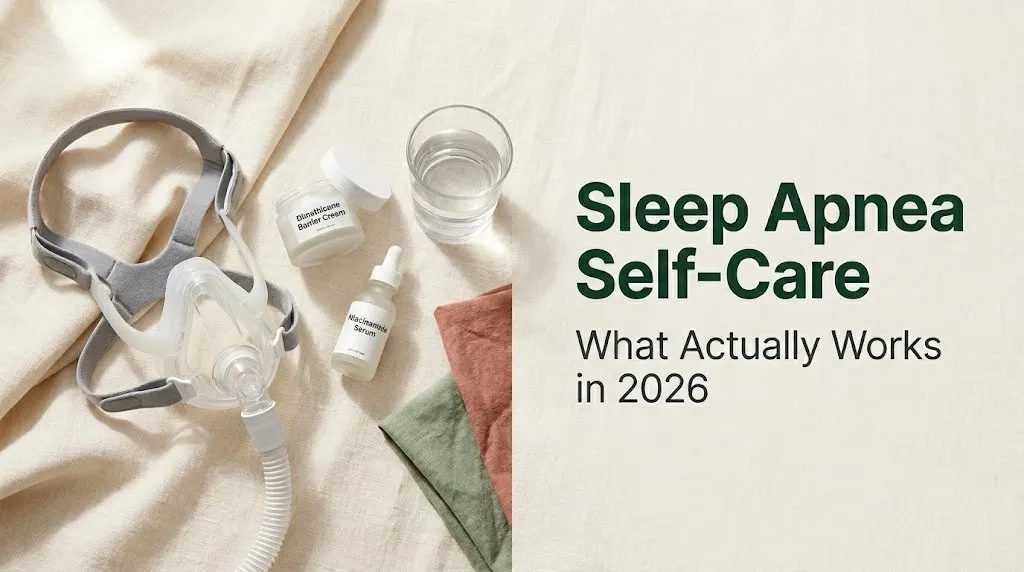
CPAP Mask Skin Irritation and How to Protect Your Skin
CPAP mask skin irritation is a documented clinical reality that affects between 30% and 50% of regular CPAP users to some degree, ranging from mild redness at pressure points to frank contact dermatitis and pressure-related skin breakdown.
The mechanisms are distinct and worth understanding separately. Pressure-related skin changes occur at the nose bridge, upper lip, and cheekbones where mask headgear creates sustained mechanical loading against the skin. Sustained pressure impairs capillary blood flow, and over hours of nightly use, this creates localized erythema (redness), micro-abrasion, and, in people with Fitzpatrick types IV through VI, a heightened risk of post-inflammatory hyperpigmentation (PIH) at pressure sites. PIH occurs when inflammatory signals from repeated tissue stress activate melanocytes to increase melanin production, even after the primary inflammation resolves.
Contact dermatitis from mask materials is a separate issue. Most CPAP mask cushions are made from medical-grade silicone. True silicone allergy is rare but documented. More commonly, the reaction is irritant contact dermatitis from the combination of silicone material, facial oils, sweat, and the physical friction of the mask moving during sleep. A 2019 report in the British Journal of Dermatology noted that silicone medical device contact dermatitis is frequently misclassified as allergic when patch testing to silicone components is negative, confirming an irritant rather than allergic mechanism in most cases.
Signs of mask-related skin irritation to watch for:
- Persistent redness lasting more than 30 minutes after removing the mask
- Papular or vesicular (blister-like) reaction along the mask contact edge (more indicative of allergic contact dermatitis than irritant type)
- Thickened or lichenified skin at chronic pressure sites
- Hyperpigmented marks at nose bridge or cheekbones that persist between mask removal and next-night use
- Broken or eroded skin at any pressure point
If vesicular or spreading rash develops, or if skin breakdown does not resolve with barrier protection within two weeks, a board-certified dermatologist should be consulted. The dermatologist may perform patch testing to specific silicone components and CPAP mask materials and can prescribe a short course of topical corticosteroid to resolve active contact dermatitis.
Skincare Routine for CPAP Users
A targeted skincare routine for nightly CPAP users focuses on three goals: protecting the skin barrier at mask contact points, preventing and managing PIH at chronic pressure sites, and keeping mask surfaces clean enough to preserve both mask function and skin health.
The evening routine (before mask application) should follow this sequence:
- Cleanse: use a gentle, non-foaming cleanser or a micellar water to remove daytime sebum, sunscreen, and any skincare residue from the skin surface. Residue on the skin transfers to the mask cushion, degrading silicone and contributing to irritant contact dermatitis.
- Treat active PIH (if present): apply a targeted hyperpigmentation treatment to pressure-point areas. Azelaic acid (at concentrations of 15% to 20%) has evidence for reducing post-inflammatory hyperpigmentation without the irritation risk of retinoids or the photosensitivity concern of high-concentration alpha-hydroxy acids. Niacinamide (INCI: niacinamide; also known as nicotinamide) at 4% to 10% inhibits melanosome transfer from melanocytes to keratinocytes, reducing PIH formation. Apply either to the nose bridge and cheekbone areas specifically.
- Apply a non-comedogenic barrier product to pressure points: a dimethicone-based barrier cream (INCI: dimethicone, CAS 9016-00-6) applied as a thin layer to the nose bridge, upper lip, and cheekbones creates a low-friction interface between skin and mask. Dimethicone does not degrade silicone mask surfaces the way petroleum-based oils can, making it the preferred choice for CPAP users over petrolatum applied directly to sealing surfaces.
- Skip any oil-based product on the mask sealing zones: oils (whether from argan, jojoba, rosehip, or any other source) degrade medical-grade silicone over time, compromise the airtight seal, and worsen both mask leak and skin irritation from vibration.
- Allow all skincare products to fully absorb before placing the mask: a 5-minute wait after applying barrier products reduces the likelihood of product migrating onto the mask sealing surface.
| Skincare Step | Product Type | Mask-Compatible? | Notes |
|---|---|---|---|
| Cleanse | Gentle non-foaming cleanser | Yes | Essential pre-mask step |
| Treat PIH | Niacinamide 4-10% or azelaic acid 15-20% | Yes (absorbs before masking) | Apply specifically to pressure zones |
| Protect barrier | Dimethicone barrier cream | Yes | Avoid on sealing surfaces |
| Moisturize | Water-based gel moisturizer | Yes if fully absorbed | Humectant-forward, minimal oil |
| Avoid | Any oil-based product on seal zones | No | Degrades silicone |
Morning routine (after mask removal):
Cleanse again to remove any mask residue and overnight sebum buildup. Follow with a gentle, hydrating toner or essence containing sodium hyaluronate (the sodium salt of hyaluronic acid, smaller molecular weight than hyaluronic acid, penetrates to the upper layers of the epidermis) to restore hydration to skin that may have experienced some overnight dehydration. Apply SPF 30 or higher broad-spectrum sunscreen to all areas, including nose bridge and cheekbones, since both azelaic acid and niacinamide make these areas more susceptible to UV-triggered PIH if photoprotection is skipped.
Key Takeaway: Dimethicone barrier cream at CPAP mask pressure points, applied before the mask, and niacinamide or azelaic acid on established PIH are the two most evidence-aligned skincare strategies for regular CPAP users. Oil-based products on the silicone sealing surface are a mask-damaging mistake worth knowing before you make it.
Sleep Apnea, Stress, and Mental Health Self-Care
Stress and sleep apnea have a bidirectional relationship that most self-care guides ignore. OSA causes stress through sleep deprivation, mood dysregulation, and the constant physiological stress of nocturnal hypoxia. Chronic stress, in turn, disrupts sleep architecture in ways that worsen OSA severity.
The mechanism runs through the hypothalamic-pituitary-adrenal (HPA) axis. Chronic psychological stress elevates cortisol, which delays sleep onset, reduces slow-wave sleep duration, and increases arousal frequency during the night. In people with OSA, more arousals mean more perceived apnea events and less restorative sleep per hour spent in bed. A 2020 review in the Journal of Sleep Research found that self-reported stress levels were independently predictive of OSA symptom severity after controlling for AHI, suggesting that stress amplifies the subjective experience of OSA beyond what the AHI alone predicts.
Stress reduction practices with evidence-informed mechanisms for sleep include:
- Mindfulness-based stress reduction (MBSR): an 8-week structured program with RCT evidence for reducing cortisol, improving sleep onset latency, and reducing nighttime arousal frequency
- Progressive muscle relaxation (PMR): activates the parasympathetic nervous system, reducing sympathetic arousal at bedtime; a 2018 meta-analysis in the Journal of Behavioral Sleep Medicine found PMR consistently reduced subjective insomnia severity and sleep onset time
- Diaphragmatic breathing: slow, controlled breathing (4 to 6 breath cycles per minute) before sleep activates the vagal brake, shifting the autonomic balance toward parasympathetic dominance; this directly counteracts the sympathetic hyperarousal pattern chronic stress creates
Depression and anxiety co-occur with OSA at rates far above the general population. According to the American Psychological Association, people with untreated OSA have approximately double the risk of clinically significant depressive symptoms compared to matched controls without OSA. CPAP treatment reduces depressive symptom scores in many but not all patients. If depressive or anxiety symptoms persist after 8 to 12 weeks of adequate CPAP use and behavioral self-care, evaluation by a licensed mental health professional (a licensed clinical psychologist or psychiatrist for medication assessment) is appropriate and warranted.
Daytime Fatigue Management for People With Sleep Apnea
Daytime fatigue in OSA is not simply a product of sleep quantity. It persists because OSA fragments the architecture of sleep itself, reducing the proportion of slow-wave sleep (the restorative deep sleep stage) and rapid eye movement (REM) sleep that the brain and body require for cellular repair, memory consolidation, and hormonal regulation.
Managing daytime fatigue requires a parallel track: improving nighttime sleep quality through all the self-care practices already discussed, while implementing specific daytime strategies that reduce the functional impact of residual fatigue.
Strategic napping (when appropriate) can reduce OSA-related cognitive impairment without worsening nighttime sleep, if limited to 20 to 30 minutes before 2 p.m. Naps longer than 30 minutes risk entering slow-wave sleep, which creates sleep inertia (the groggy, disoriented feeling on waking) and reduces homeostatic sleep pressure enough to delay nighttime sleep onset.
Physical activity timing matters for daytime energy. A 2020 study in the Annals of the American Thoracic Society found that moderate-intensity exercise performed in the morning or early afternoon improved OSA-related daytime sleepiness more than the same exercise performed in the evening, where it slightly delayed sleep onset. Morning exercise also provides light exposure that strengthens the circadian wake signal, supporting the consistent wake time that anchors sleep hygiene.
Caffeine management is a practical lever. Caffeine has a half-life of 5 to 7 hours in most adults (longer in people who metabolize caffeine slowly due to CYP1A2 gene variants). Consuming caffeine after noon, in a person who sleeps at 10 to 11 p.m., means a quarter of that caffeine remains active at bedtime, worsening sleep onset latency and reducing slow-wave sleep time.
People with OSA who experience daytime sleepiness severe enough to impair driving or occupational performance should discuss this specifically with their sleep medicine physician. The physician may assess whether wakefulness-promoting medications (armodafinil or modafinil, FDA-approved for OSA-related excessive daytime sleepiness) are appropriate as an adjunct to CPAP and behavioral self-care.
When Sleep Apnea Self-Care Is Not Enough: Professional Care Thresholds
Sleep apnea self-care is never a substitute for professional evaluation and, where indicated, medical treatment. Knowing the specific clinical thresholds that require professional escalation is as important as knowing which self-care practices to prioritize.
The American Academy of Sleep Medicine specifies these clinical thresholds clearly:
- AHI of 15 or above (moderate OSA): positive airway pressure therapy is indicated regardless of symptoms. Self-care can be adjunctive but does not meet the standard of care as sole treatment.
- AHI of 5 to 14 with symptoms (mild symptomatic OSA): treatment is indicated; options include CPAP, oral appliance therapy from a dentist trained in dental sleep medicine, or positional therapy with monitoring. Self-care alone requires close follow-up.
- AHI of 5 or above with cardiovascular comorbidities: the American Heart Association’s 2021 scientific statement on OSA and cardiovascular disease states that any level of OSA with co-occurring hypertension, heart failure, atrial fibrillation, or stroke warrants CPAP evaluation regardless of AHI severity classification.
Anyone who has not had formal sleep testing (polysomnography or a validated home sleep apnea test) and is managing presumed OSA entirely through self-care needs to understand that AHI cannot be estimated from symptoms alone. Daytime sleepiness, snoring, and witnessed apnea are suggestive but not diagnostic. A board-certified sleep medicine physician can order and interpret a sleep study, determine AHI, and confirm whether self-care is appropriate as primary management or adjunctive to prescribed therapy.
Self-care must also be escalated to professional care when:
- Daytime sleepiness impairs driving, occupational safety, or daily functioning
- Blood pressure remains elevated despite adequate CPAP use and lifestyle modification
- New cardiac symptoms (palpitations, chest discomfort, shortness of breath) develop
- Mood disturbance (depression or anxiety) persists after 8 to 12 weeks of consistent CPAP use and self-care
The reader who takes self-care seriously and tracks their response honestly, whether through CPAP data, sleep diary, daytime symptom logging, or home monitoring, gives their sleep medicine physician the most useful data for adjusting treatment appropriately.
Frequently Asked Questions About Sleep Apnea Self-Care
Can sleep apnea go away on its own with lifestyle changes?
Sleep apnea rarely resolves entirely on its own without targeted intervention, but lifestyle changes can meaningfully reduce severity in mild to moderate cases.
A 10% body weight reduction is associated with a 26% average AHI reduction, per research published in the Archives of Internal Medicine, and oropharyngeal exercises have reduced AHI by approximately 39% in randomized controlled trials.
Whether AHI drops below the diagnostic threshold of 5 events per hour depends on the individual’s baseline severity, anatomical factors, and the consistency of their self-care practice.
What sleeping position is best for sleep apnea?
Sleeping on your side (lateral decubitus position) is the most evidence-supported sleeping position for reducing OSA severity.
According to data published in the Journal of Clinical Sleep Medicine, 50% to 60% of OSA cases are positional, meaning the supine AHI is at least double the lateral AHI.
Head-of-bed elevation by 30 to 45 degrees also reduces AHI and is particularly useful for people who also experience acid reflux.
Does alcohol make sleep apnea worse?
Yes, alcohol consistently worsens OSA severity by depressing hypoglossal nerve function and reducing genioglossus muscle activation during sleep.
A 2018 analysis in the Journal of Sleep Research found that alcohol before bed increased AHI by a mean of 25% compared to alcohol-free nights.
Avoiding alcohol within 3 to 4 hours of sleep is the minimum recommended adjustment for people with any severity of OSA.
How long does it take for self-care to improve sleep apnea symptoms?
Subjective sleep quality improvements from behavioral changes often appear within 2 to 4 weeks, but measurable AHI reductions require 8 to 12 weeks of consistent practice.
Positional therapy produces immediate AHI changes on the first night of correct implementation in people with positional OSA.
Oropharyngeal exercises require 3 months of daily practice to achieve the 39% AHI reduction documented in randomized controlled trials.
Can CPAP masks cause skin problems?
Yes, between 30% and 50% of regular CPAP users experience some degree of mask-related skin irritation, ranging from pressure redness to contact dermatitis.
The most common cause is irritant contact dermatitis from the combination of silicone material, facial oils, sweat, and mask friction.
People with Fitzpatrick skin types IV through VI face an additional risk of post-inflammatory hyperpigmentation at chronic pressure points, which responds to niacinamide at 4% to 10% or azelaic acid at 15% to 20%.
Is sleep apnea self-care safe for people with severe OSA?
Self-care practices are safe and beneficial as adjunctive habits for people with severe OSA, but they must not replace prescribed CPAP or bilevel PAP therapy.
The American Academy of Sleep Medicine specifies that severe OSA (AHI above 30) requires positive airway pressure therapy regardless of the outcome of lifestyle modifications.
Anyone with severe OSA managing the condition without CPAP should discuss this directly with a board-certified sleep medicine physician, who can clarify the cardiovascular risk of untreated severe OSA.
Closing
The self-care practices that genuinely move the needle on sleep apnea share one characteristic: they work on a specific, named physiological mechanism. Weight loss narrows parapharyngeal fat. Lateral positioning uses gravity correctly. Oropharyngeal exercises strengthen the muscles that keep the airway open. Alcohol avoidance protects hypoglossal nerve function. Each of these has real, named clinical trial evidence. They are not wellness trends.
If you are a CPAP user, the nightly skincare step is not optional maintenance. It is active protection of skin that takes 8 or more hours of mechanical contact per night. Dimethicone barrier cream at pressure points and niacinamide or azelaic acid for any established PIH are specific, achievable, and evidence-aligned additions to your existing routine.
Start with the two changes with the strongest evidence and the lowest barrier: consistent wake time every morning and no alcohol within 3 to 4 hours of sleep. Track how you feel over 4 weeks. Then add positional therapy or oropharyngeal exercises. You now have a clearer picture of the evidence than most people, and that clarity is where sustainable change actually begins.